What to do in an emergency? How you can help
5. August 2025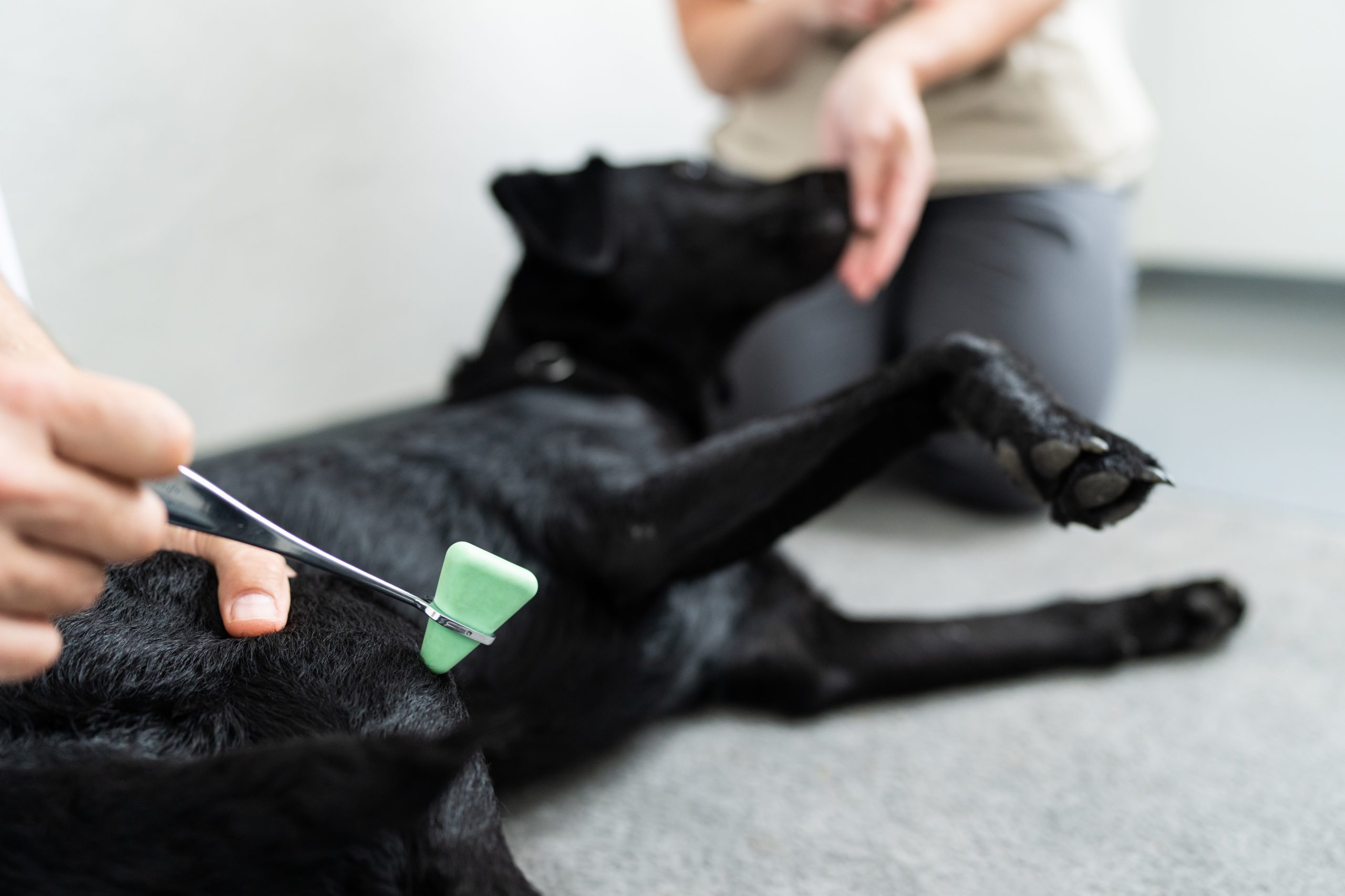
Animal neurology: What you should know
5. August 2025What to do in an emergency? How you can help
5. August 2025Animal neurology: What you should know
5. August 2025Arthroscopy in Animals: A Look Inside the Joint
Arthroscopy, also known as joint endoscopy, is a modern, minimally invasive procedure for the examination and treatment of joint problems in animals. Using a small camera called an arthroscope, we can look directly into the affected joint without making large incisions. This allows us to identify damage precisely and treat it in a targeted manner.
What is Arthroscopy?
Minimally invasive arthroscopy has proven to be a precise and joint-preserving method in orthopedic diagnostics and surgery. It enables us to detect joint problems early and, if necessary, to intervene therapeutically during the procedure.
When is Arthroscopy Used?
Arthroscopy is used for many orthopedic problems, particularly for:
-
Cruciate ligament problems
-
Osteochondrosis dissecans (OCD)
-
Meniscus injuries
-
Free joint bodies
-
Joint inflammation (arthritis)
-
Misalignments
- Ligament tears
- Osteoarthritis
All joints can be arthroscoped safely and effectively.
Why Minimally Invasive? Advantages of Modern Joint Endoscopy
Compared to traditional open joint surgery, arthroscopy offers many advantages:
-
Minimally invasive: No large incision, resulting in less pain and lower risk of complications
-
Better visualization using 4K/HD camera
-
Rapid recovery: Animals are usually mobile again after just a few days
-
Reduced scarring: Only minimal skin openings required
-
Lower infection risk: Less tissue trauma means less risk of inflammation
-
Direct therapy during examination: Damage can be treated immediately
-
Gentle on the joint: No unnecessary trauma to surrounding structures
With this technique, we can not only accurately assess the condition of the joint but also intervene therapeutically immediately, e.g., remove inflamed tissue or perform joint lavage.
How Arthroscopy Works: Step by Step
-
Preliminary examination: Detailed lameness diagnostics, X-rays, and CT scans beforehand
-
Anesthesia induction: Arthroscopy is always performed under general anesthesia
-
Insertion of the arthroscope: Tiny incision, view into the joint through a high-resolution camera – particularly gentle and minimally invasive thanks to our 1.1 mm Nanoscope system.
-
Diagnostics & Therapy: Depending on findings, surgical measures are implemented immediately (e.g., removal of free joint bodies, joint lavage, smoothing of cartilage damage)
-
Wound closure & Aftercare: Small wounds, short healing process
Aftercare: What Happens After Arthroscopy?
-
Animals may walk again on the same day or the next day
-
You will receive a clear aftercare plan with medications, rest period, and physiotherapy if necessary
-
Follow-up examination to monitor progress typically after approximately 10–14 days
-
Our team is available for aftercare or additional treatments if needed
Modern Veterinary Medicine for Better Quality of Life
Especially with joint problems, early diagnosis is crucial to prevent secondary damage and improve your animal's quality of life. With arthroscopy, we offer you a modern, gentle, and effective solution for treating orthopedic conditions.
Many joint diseases can be detected at a young age. Arthroscopic examination is possible, for example, at:
-
Hip: approximately 16–20 weeks
-
Elbow: from approximately 4–5 months
-
Shoulder: from approximately 5–6 months
Conclusion: Precise Help for Joint Problems: Gentle and Effective
Arthroscopy is a milestone in surgical veterinary medicine. It enables early diagnosis, direct therapy, and faster healing, while placing minimal stress on your animal.
Whether lameness, chronic conditions, or acute joint problems:
The earlier you act, the better the chances of recovery. With arthroscopy, we offer a gentle, modern, and effective solution so your animal can move through life with joy again.
